Plagiocephaly
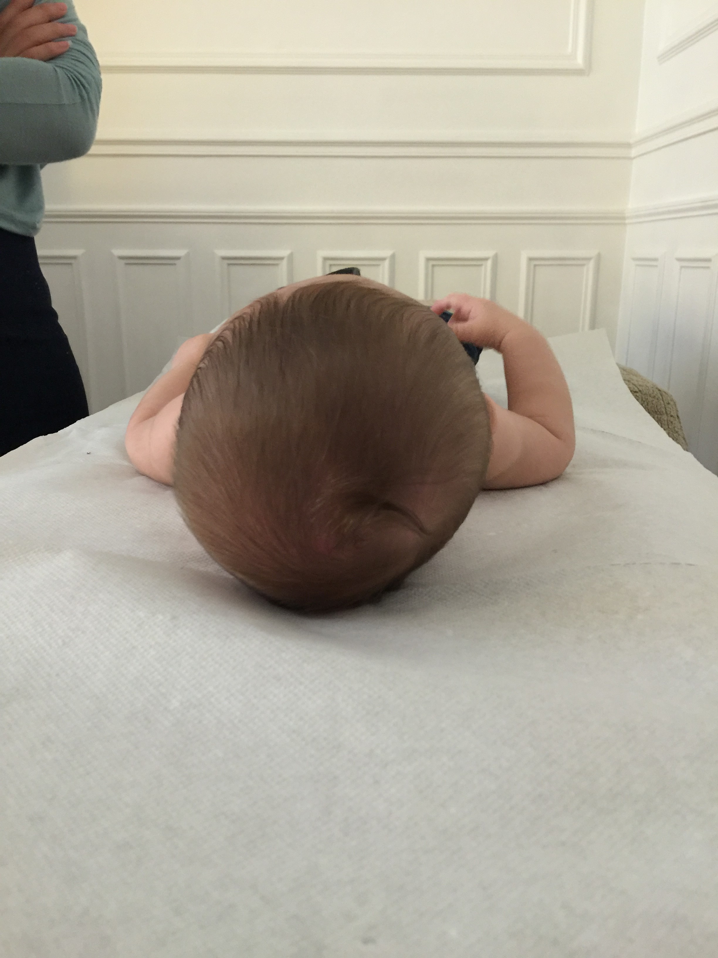
Plagiocephaly is a relatively common lesion characterized, most commonly, by a flattening of the posterior part of the skull on the right side.

The asymmetrical skull of plagiocephaly, seen from above.
There are two forms:
- Synostotic plagiocephaly due to the closure of one or more cranial sutures. It will probably be treated surgically and is fortunately very rare. The mere fact of its existence will require a proper medical diagnosis…
- The so-called positional plagiocephaly that is classically attributed to the infant’s bed. This is the one we will talk about here.
For an infant with a flattening of the posterior part of the skull on the right side.
This deformity appears to be associated with a torticollis related to a “spasm” of a neck muscle called Sterno Cleido Occipito Mastoid (SCOM). This spasm will most frequently be located on the left side. Also the infant will have difficulty turning his head to the right.
First of all don’t panic, this infant will not keep this oblong form so marked all his life, even if he is not treated. But he will suffer all the same, on the one hand because the fact of having a torticollis permanently should not be very pleasant for him and on the other hand other invisible lesions will make the bed of other symptoms, present or future.
The first thing to do is to reassure the parents. Normally, with very rare exceptions, everything is going to be fine.
But first of all, why does one become “plagiocephalic” and how?

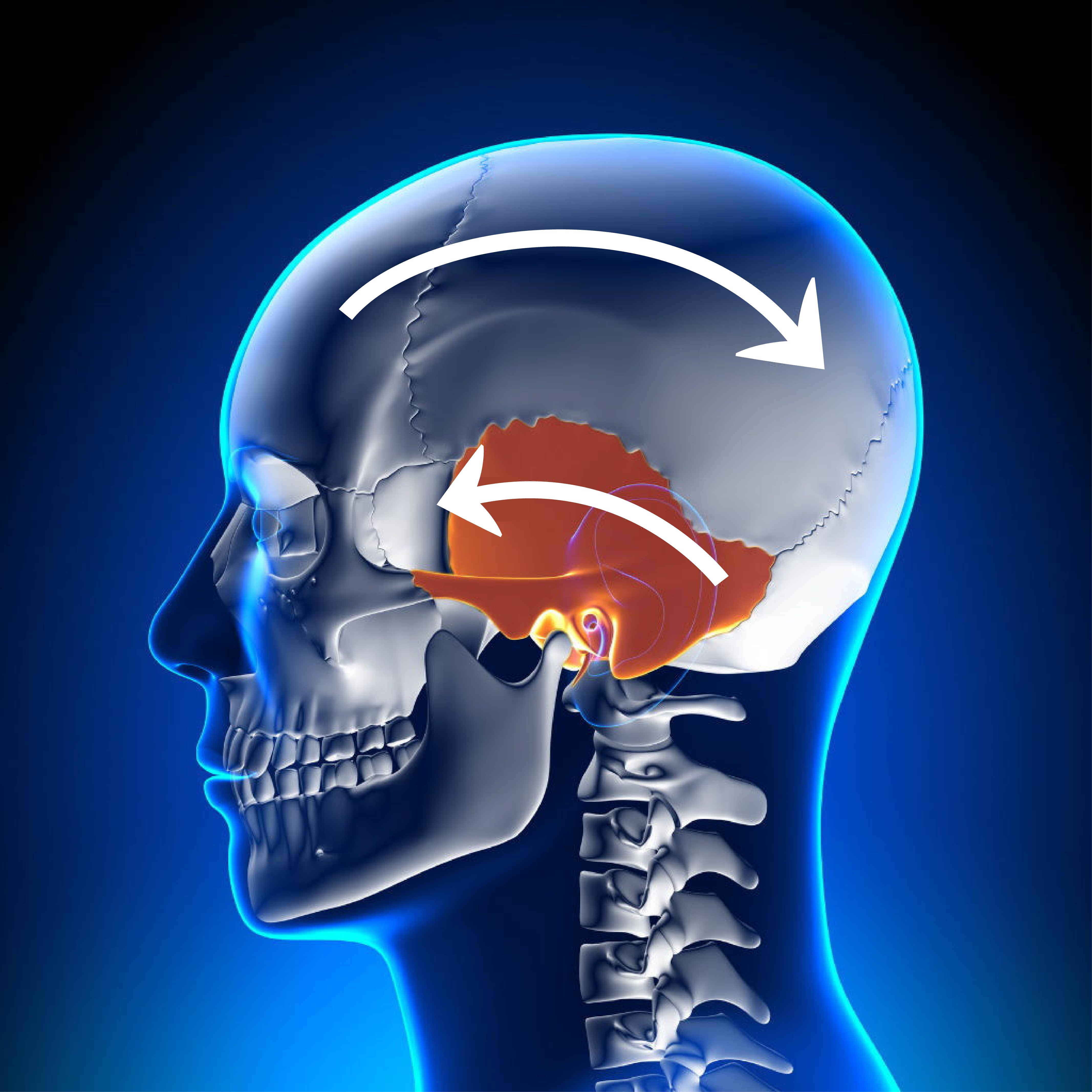
The movements of the skull bones that we perceive during thoracic inspiration.
A little anatomy
In this illustration of a human skull you see the temporal located in the center. What we can feel are the movements of the temporal in relation to the other bones of the skull.
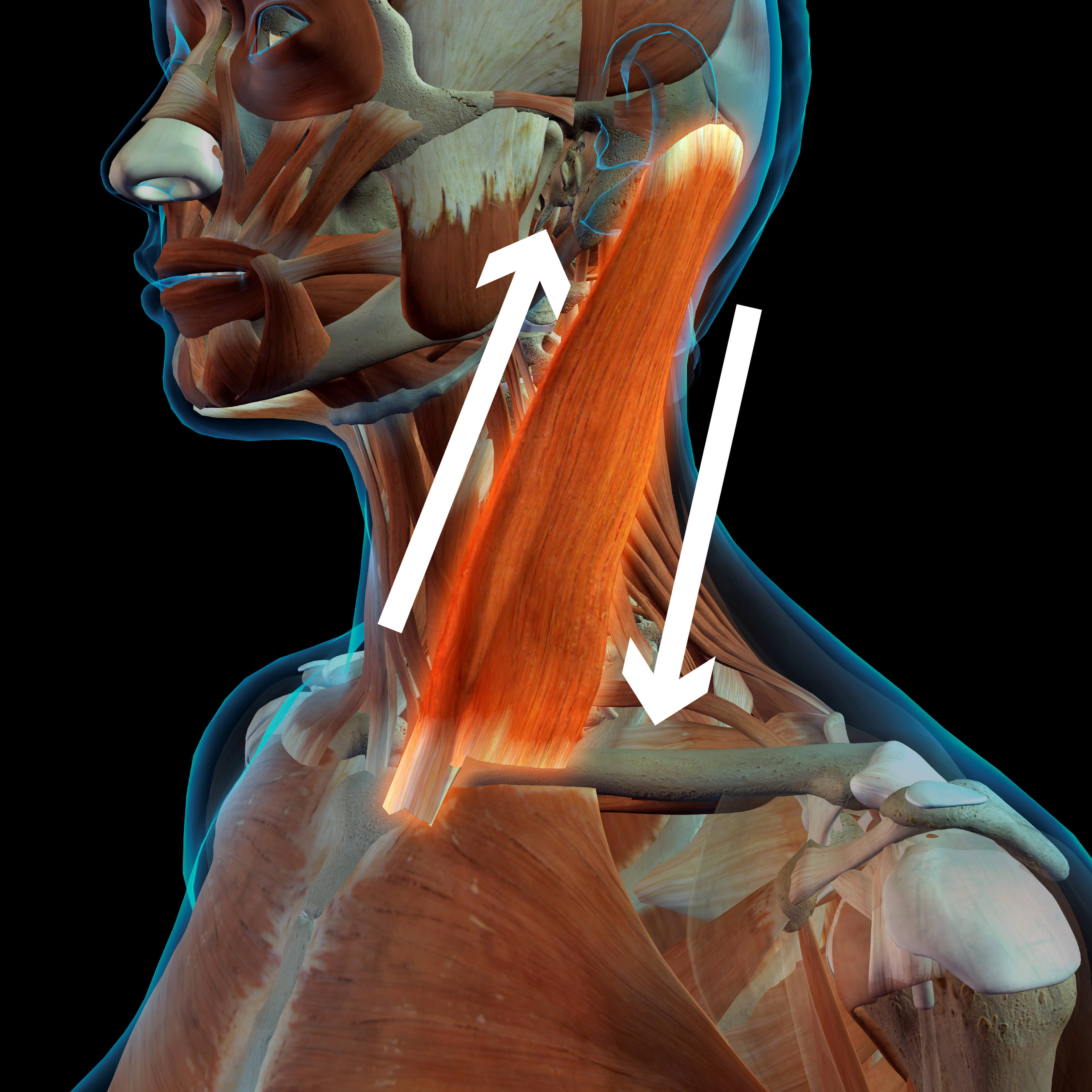
The Sterno Cleido Occipito Mastoidian muscles, which are inserted at the top of the temporals and at the bottom of the sternum and clavicles, will be the origin of (almost imperceptible) movements that will mobilize the temporals in relation to the other bones of the skull. The two anterior bundles contract while the two posterior bundles relax and vice versa. And this in an alternating and permanent way, even at rest. The body needs these micro-movements permanently in order to drain the tissues and organs.

The anterior muscles will bring the temporal in anterior rotation, the posterior muscles in posterior rotation. Their contractions will be alternated.
These muscles will connect the temporal bones to the first dorsal vertebra via the clavicles.
Normally the two temporal bones will have symmetrical movements in relation to the other bones of the skull. But there are lesions that reverse the movements of one of the two temporal bones. We call these lesions “desynchronization lesions”.
A desynchronization injury occurs when an individual goes into extreme rotation, and brings his head back to the neutral position while exhaling (which is anti physiological). The first dorsal vertebra associated with the clavicle will be a better candidate, which will be out of sync, causing an (osteopathic) lesion of the temporal vertebrae that causes plagiocephaly.
Thus, while one temporal will go in anterior rotation, the other will go in posterior rotation. The skull will then function in torsion and will become deformed. If in an adult a temporal torsion can go unnoticed, it is not necessarily the same in an infant (but it is possible).
It is because the lesion is produced by bringing the head back to the neutral position and exhaling that we can say that lying on one’s back is more conducive to plagiocephaly. Obviously, lying on one’s stomach excludes this lesion mechanism…
In adults, treatment is done by turning the patient’s head away from the temporal side of the lesion until mobilization of the clavicle is initiated. Then the patient is asked to make a respiratory apnea in inspiration, to bring the head back by turning it to the neutral position and to blow. The two temporals will then be synchronous again.
But then for infants, the situation seems to be more complicated, as they systematically refuse to comply with the inspiratory apnea. So we have to adapt our technique and here’s how… But before doing so, be careful to note that the child turns his head less well to one side than the other (especially to the right). This will be your reference to check if your technique has been effective.
The infant is lying on his back. In the role of the therapist we will take, for example, the father and in the role of object of covetousness we will take, if possible, the mother. But you will be able to redistribute the roles according to your convenience. The Mother is placed on the side where the infant turns his head most easily. Theoretically it will be on the side where the neck muscle (SCOM) is the most contracted, i.e. most often on the left. The therapist (Father) places one hand on the infant’s left cheek (on the Mother’s side) and the other hand on the opposite shoulder (right) of the infant.
Ask the Mother to lower her head to the table plane to encourage the child to turn his head as much as possible until the therapist feels that the rotation of the child’s head moves his right shoulder (the infant’s collarbone) under his (the Father’s) hand. Once the rotational movement of the child’s head is felt by the Father’s hand, the Father will wait for the child to breathe in (he feels it with the hand on the infant’s chest) and then (gently) return the infant’s head to the neutral position.
It is as simple as that…
You may not believe it, try it.
If all the gestures have been done correctly, the child will start to turn his head almost instantaneously much more easily to the other side. This will be the best test for someone who does not have an educated hand in the art of palpation. The Mother will go to the other side of the table to induce the child to turn his head to the other side, to verify the effectiveness of the technique. The child will then turn his head towards her much more easily.
These maneuvers give good results, and consistently.
Afterwards the child will be calmer (he will be rid of a stiff neck that has gone unnoticed), the neck muscles will gradually relax. The physiological movements of the skull, now restored in their symmetry, will help to reshape the infant’s skull instead of bringing it into torsion.
The mechanical physiological processes having returned to normal, a simple therapeutic supervision should suffice. Indeed the child could become out of sync again; it would be sufficient to start the technique again.
Obviously the skull does not regain a harmonious shape with the wave of a magic wand. It will still take quite some time for it to return to a quasi-symmetry, but it has passed from a lesional phase to a normal physiological phase (of self-healing).
Other so-called cranial osteopathic techniques will eventually give the infant’s skull a more fluid mobility, thus accelerating the process of self-remodelling of the skull. But we do not have the necessary hindsight to say so. Infants whose temporal muscles have just been synchronized are generally more relaxed after the treatment.
There are many plagiocephalic children whose diagnosis is not established because the deformity is not obvious to the lay eye. The link between a form of infant torticollis and plagiocephaly seems obvious to us. When examining the rotation of the infant’s head, we will often find loss of amplitude on one side which will allow us to detect a slight deformation of the child’s skull.
There are many plagiocephalies that go unnoticed.
Be vigilant.
Helping infants around the world
You can quickly store and share this folder by downloading it in English or French with the following links:
